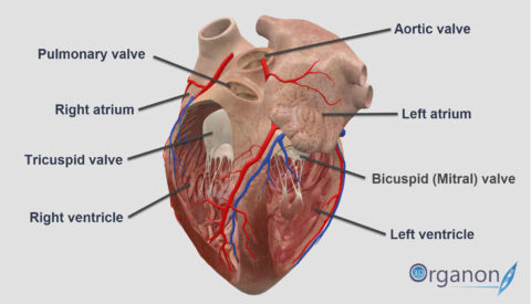
42 the human heart and its labels
Human Heart - Anatomy, Functions and Facts about Heart The human heart is divided into four chambers, namely two ventricles and two atria. The ventricles are the chambers that pump blood and atrium are the chambers that receive the blood. Among which, the right atrium and ventricle make up the "right portion of the heart", and the left atrium and ventricle make up the "left portion of the heart." 5. 13 parts of the human heart (and its functions) - LORECENTRAL Its opening (generated by the systole of the atrium) causes blood to travel between both regions. 3. Left Ventricle. Another major part of the heart. The left ventricle receives oxygen-rich blood from the left atrium and sends it to the rest of the body through the aortic artery. 4. Aortic sigmoid valve.
› US › enAC16 Human Cardiomyocyte Cell Line | SCC109 - EMD Millipore AC16 Human Cardiomyocyte Cell Line is a proliferating cell line that was derived from the fusion of primary cells from adult human ventricular heart tissues with SV40 transformed, uridine auxotroph human fibroblasts, devoid of mitochondrial DNA (1, 2).

The human heart and its labels
Diagram of Human Heart and Blood Circulation in It Exterior of the Human Heart A heart diagram labeled will provide plenty of information about the structure of your heart, including the wall of your heart. The wall of the heart has three different layers, such as the Myocardium, the Epicardium, and the Endocardium. Here's more about these three layers. Epicardium Human Heart - Diagram and Anatomy of the Heart - Innerbody The heart is a muscular organ about the size of a closed fist that functions as the body's circulatory pump. It takes in deoxygenated blood through the veins and delivers it to the lungs for oxygenation before pumping it into the various arteries (which provide oxygen and nutrients to body tissues by transporting the blood throughout the body). Parts Of The Human Heart | Science Trends The parts of the human heart can be broken down into four chambers, muscular walls, vessels, and a conductive system. The two upper chambers are called the atria, with lower parts called ventricles. These all work together to make up the vital function of your heart. Everybody knows that the human heart is the essential organ in our bodies.
The human heart and its labels. Human Heart Diagram Labeled | Science Trends Human Heart Diagram Labeled Daniel Nelson 1, January 2019 | Last Updated: 3, March 2020 The human heart is an organ responsible for pumping blood through the body, moving the blood (which carries valuable oxygen) to all the tissues in the body. Without the heart, the tissues couldn't get the oxygen they need and would die. Given alongside is a diagram of the human heart showing its internal ... Given alongside is a diagram of the human heart showing its internal structure. Label the parts marked 1 to 6, and answer the following questions.Which chamb... Human heart: Anatomy, function & facts | Live Science The human heart is an organ that pumps blood throughout the body via the vessels of the circulatory system, supplying oxygen and nutrients to the tissues and removing carbon dioxide and other ... Heart Labeling Quiz: How Much You Know About Heart Labeling? Here is a Heart labeling quiz for you. The human heart is a vital organ for every human. The more healthy your heart is, the longer the chances you have of surviving, so you better take care of it. Take the following quiz to know how much you know about your heart. Questions and Answers 1. What is #1? 2. What is #2? 3. What is #3? 4. What is #4?
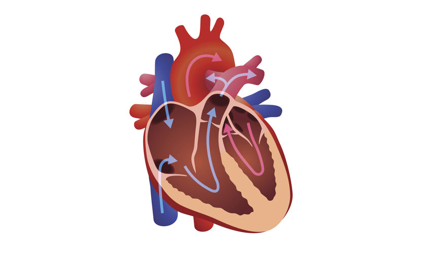
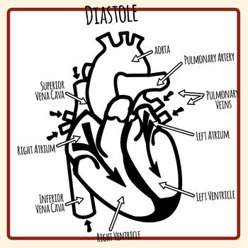
Human Heart for Kids: 2 Fun Heart Models plus Worksheets We also make a playdough heart model and completed the free printable heart worksheets. This is just one of our human body activities as part of our human body for kids unit studying anatomy for grade 1, grade 2, grade 33, grade 4, grade 5, grade 6, and garde 7 students. Whether you are a parent, teacher, or homeschooler you will love these ... A Diagram of the Heart and Its Functioning Explained in Detail The heart blood flow diagram (flowchart) given below will help you to understand the pathway of blood through the heart.Initial five points denotes impure or deoxygenated blood and the last five points denotes pure or oxygenated blood. 1.Different Parts of the Body ↓ 2.Major Veins ↓ 3.Right Atrium ↓ 4.Right Ventricle ↓ 5.Pulmonary Artery ↓ 6.Lungs Heart Diagram with Labels and Detailed Explanation - BYJUS Diagram of Heart. The human heart is the most crucial organ of the human body. It pumps blood from the heart to different parts of the body and back to the heart. The most common heart attack symptoms or warning signs are chest pain, breathlessness, nausea, sweating etc. The diagram of heart is beneficial for Class 10 and 12 and is frequently ... A Labeled Diagram of the Human Heart You Really Need to See The human heart, comprises four chambers: right atrium, left atrium, right ventricle and left ventricle. The two upper chambers are called the left and the right atria, and the two lower chambers are known as the left and the right ventricles. The two atria and ventricles are separated from each other by a muscle wall called 'septum'.
Human Heart (Anatomy): Diagram, Function, Chambers, Location in Body The heart is a muscular organ about the size of a fist, located just behind and slightly left of the breastbone. The heart pumps blood through the network of arteries and veins called the... Human Heart Labeling Teaching Resources | Teachers Pay Teachers Human Heart Parts and Blood Flow Labeling Worksheets - Diagram/Graphic Organizer by TechCheck Lessons 22 $2.25 Zip This resource contains 2 worksheets for students to (1) label the parts of the human heart and (2) Fill in a flowchart tracing the path of blood flowing though the circulatory system. Answer keys included. The Human Heart - Anatomy & Passage Of Blood - TeachPE.com The heart pumps continuously, without resting and without becoming fatigued. Its function is to pump blood to the lungs and around the body. The heart is one of the key organs in the Circulatory System. Anatomy of the heart. The heart consists of four chambers and is divided into left and right by a wall of muscle called the septum. Parts Of The Human Heart | Science Trends The parts of the human heart can be broken down into four chambers, muscular walls, vessels, and a conductive system. The two upper chambers are called the atria, with lower parts called ventricles. These all work together to make up the vital function of your heart. Everybody knows that the human heart is the essential organ in our bodies.

Congestive Heart Failure: The Essence of Heart Failure Course | CEUfast Nursing Continuing Education
Human Heart - Diagram and Anatomy of the Heart - Innerbody The heart is a muscular organ about the size of a closed fist that functions as the body's circulatory pump. It takes in deoxygenated blood through the veins and delivers it to the lungs for oxygenation before pumping it into the various arteries (which provide oxygen and nutrients to body tissues by transporting the blood throughout the body).

Something That Won't Stop Until You Die
Diagram of Human Heart and Blood Circulation in It Exterior of the Human Heart A heart diagram labeled will provide plenty of information about the structure of your heart, including the wall of your heart. The wall of the heart has three different layers, such as the Myocardium, the Epicardium, and the Endocardium. Here's more about these three layers. Epicardium

Business Diary: October 2011

30 Human Heart With Label - Labels Design Ideas 2020

New Photos in Anatomy of human body organs

Parts Of Heart Diagram Stock Illustration - Download Image Now - iStock
Our World | What is the heart?

Human Heart Pictures with Labels Best Of File Diagram Of the Human Heart Hug Wikimedia Mons ...

Simplified Heart Labeled Decal | Shop Fathead Anatomical Images Graphics
32 Label This Anterior View Of The Human Heart - Labels Design Ideas 2020
Sampoerna Wallpaper: The Heart Diagram Labeled
Human Heart - Label the Diagram Anatomy Clip Art Set Commercial Use

Pokemon Cosplay: Kawaii Pokemon Pikachu Cosplay Couple
:max_bytes(150000):strip_icc()/human-heart-circulatory-system-598167278-5c48d4d2c9e77c0001a577d4.jpg)
The Anatomy of the Heart, Its Structures, and Functions

Human Nervous System Structure and Functions Explained With Diagrams - Bodytomy
How the human heart did become associated with love? | 3D ORGANON
Post a Comment for "42 the human heart and its labels"